

- Medicine医药:奈特心脏病学彩色图谱——Marschall S.Runge E.Magnus Ohman-2007(626P):2023年-12月-16日
- Medicine医药:奈特妇产科彩色图谱——Roger P.Smith-2007(584P):2023年-12月-16日
- Medicine医药:奈特人体胚胎学彩色图谱——Larry R.Cochard-2004.PicsFolder(254P):2023年-03月-31日
- Medicine医药:奈特简明骨科学彩色图谱——Jon C.Thompson-2007(338P):2023年-03月-31日
- Medicine医药:奈特骨科疾病彩色图谱——Walter B.Greene-2009(422P):2023年-03月-31日
- Medicine医药:奈特病理学彩色图谱(528P):2023年-03月-30日
- Medicine医药:奈特人体生理学彩色图谱(218P):2023年-03月-29日
- Medicine医药:奈特药理学彩色图谱(407P):2023年-03月-29日
- Medicine医药:奈特人体神经解剖彩色图谱(309P):2023年-03月-29日
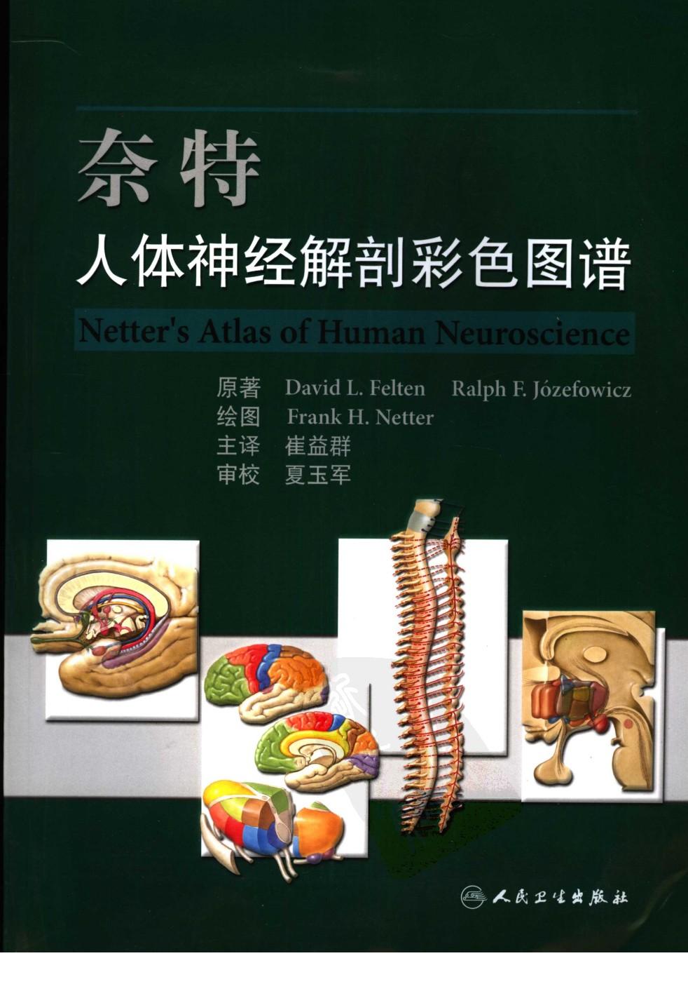
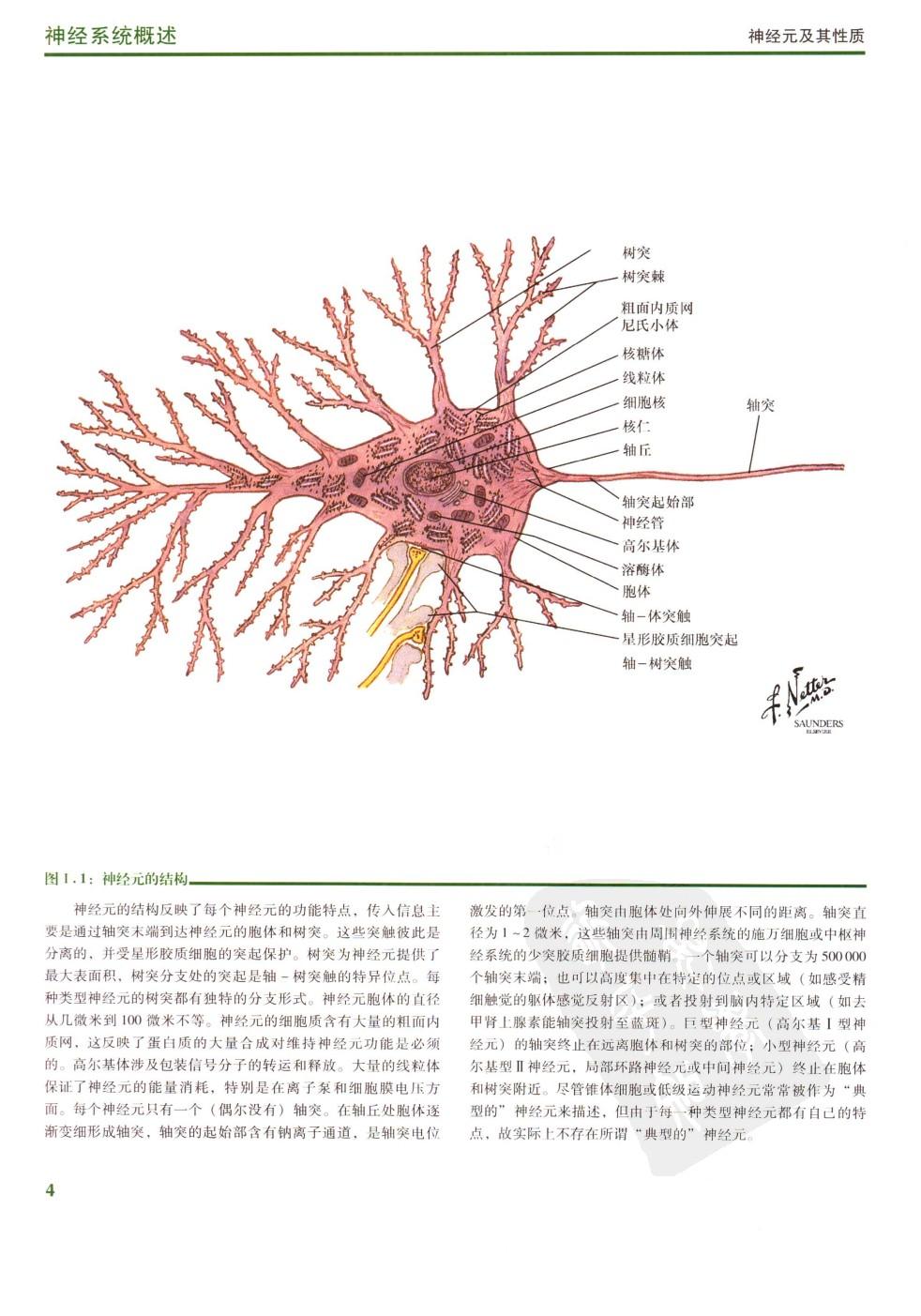
奈特人体神经解剖彩色图谱——David L.Felten Ralph F.Jozefowicz-2006,《奈特人体神经解剖彩色图谱》是2006年9月人民卫生出版社出版的图书,作者是费尔腾。本书阐述了对人类大脑、脊髓和周围神经系统许多区域的日益深入的研究成果。
《奈特人体神经解剖彩色图谱》一书,不仅涵盖Frank H.Netter博士所有原创的局部神经解剖学和系统神经解剖学丰富而精美的绘图,而且介绍了目前对人类大脑、脊髓和周围神经系统许多区域的日益深入的研究等内容,为神经学科的研究者和教师提供了该领域更广阔和更全面的知识。
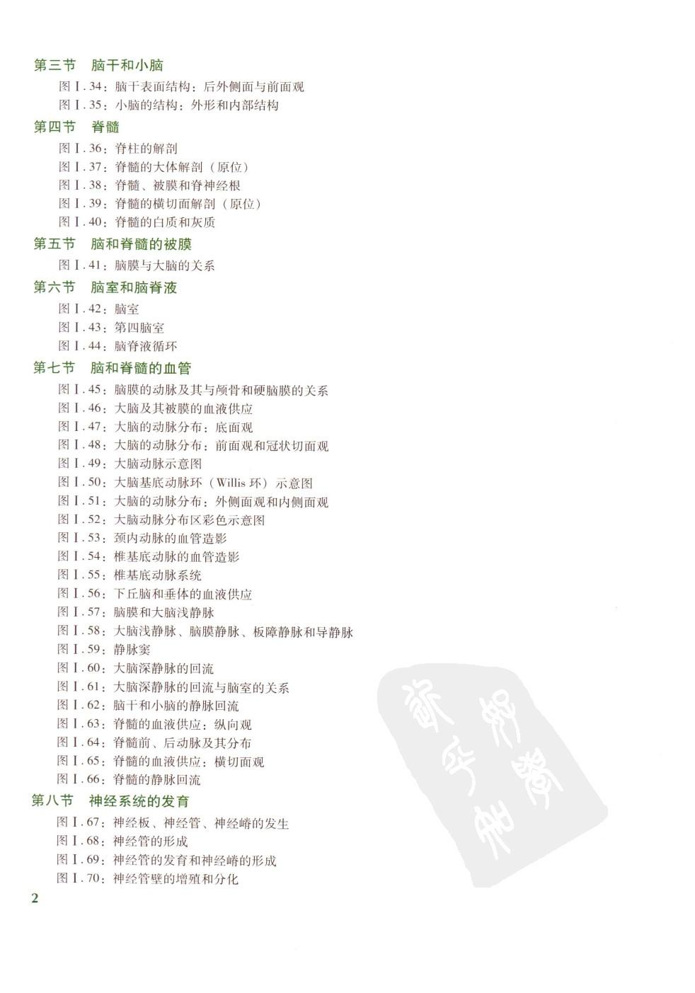
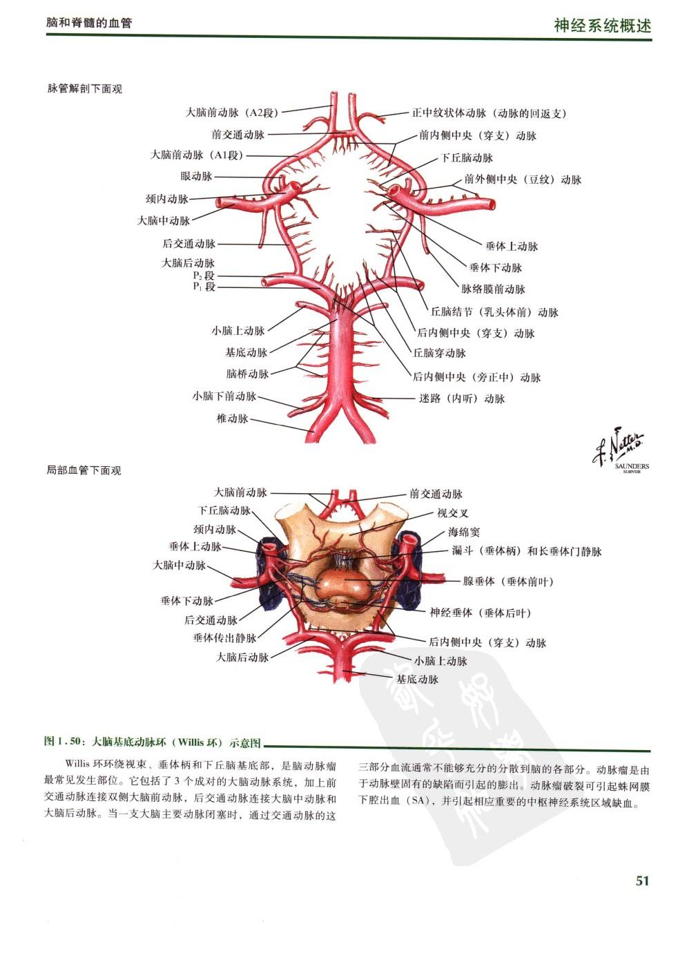
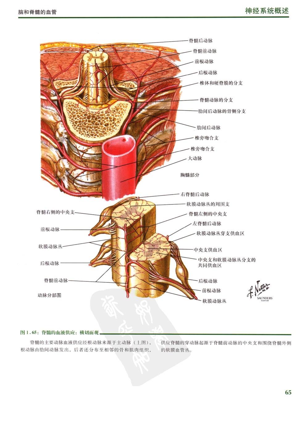
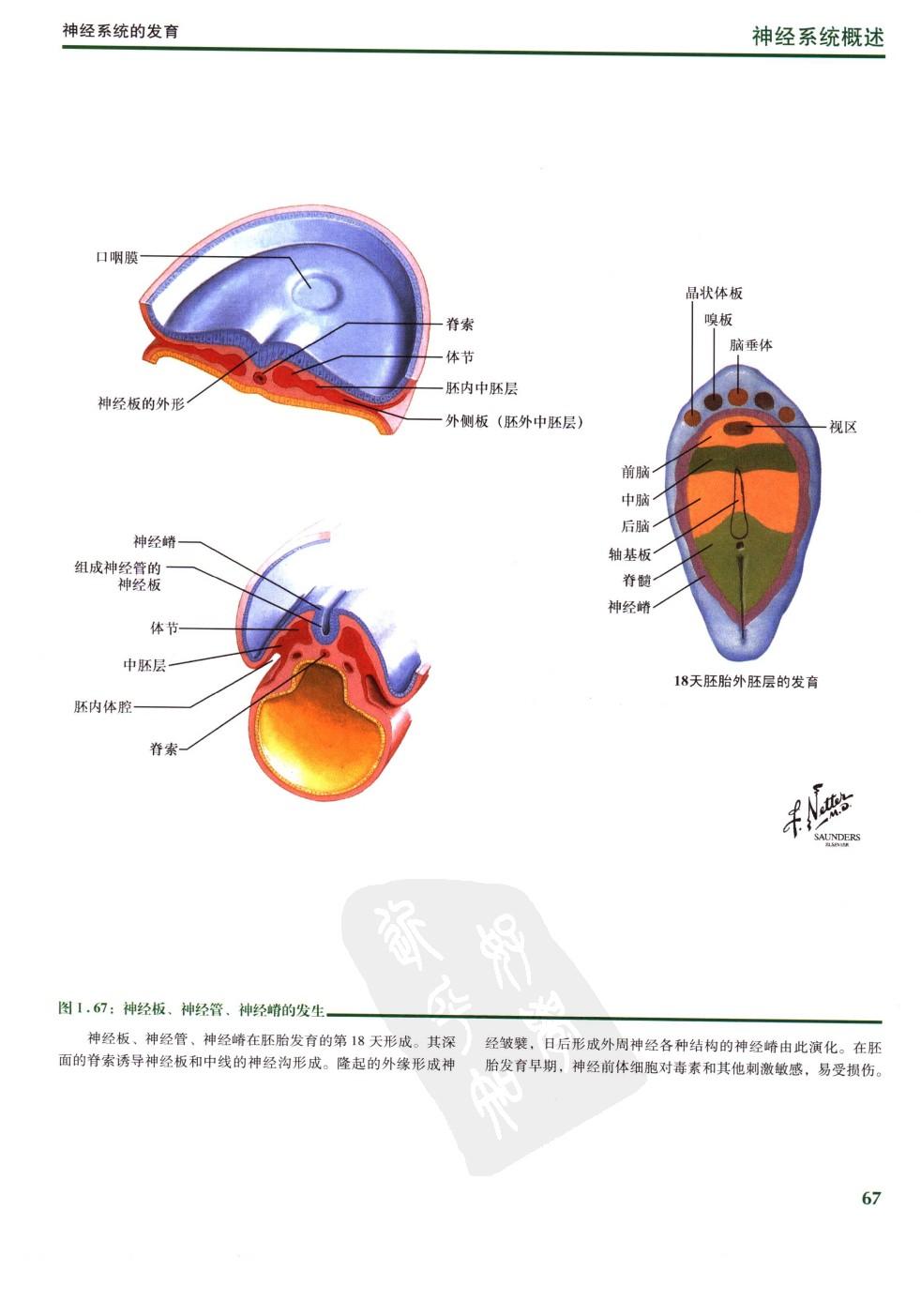
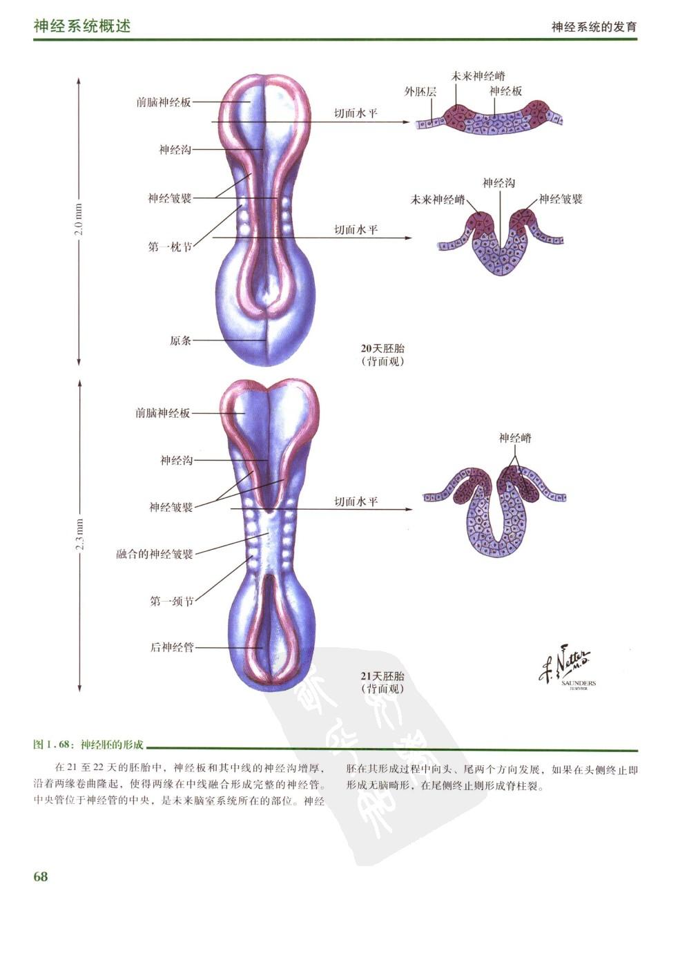
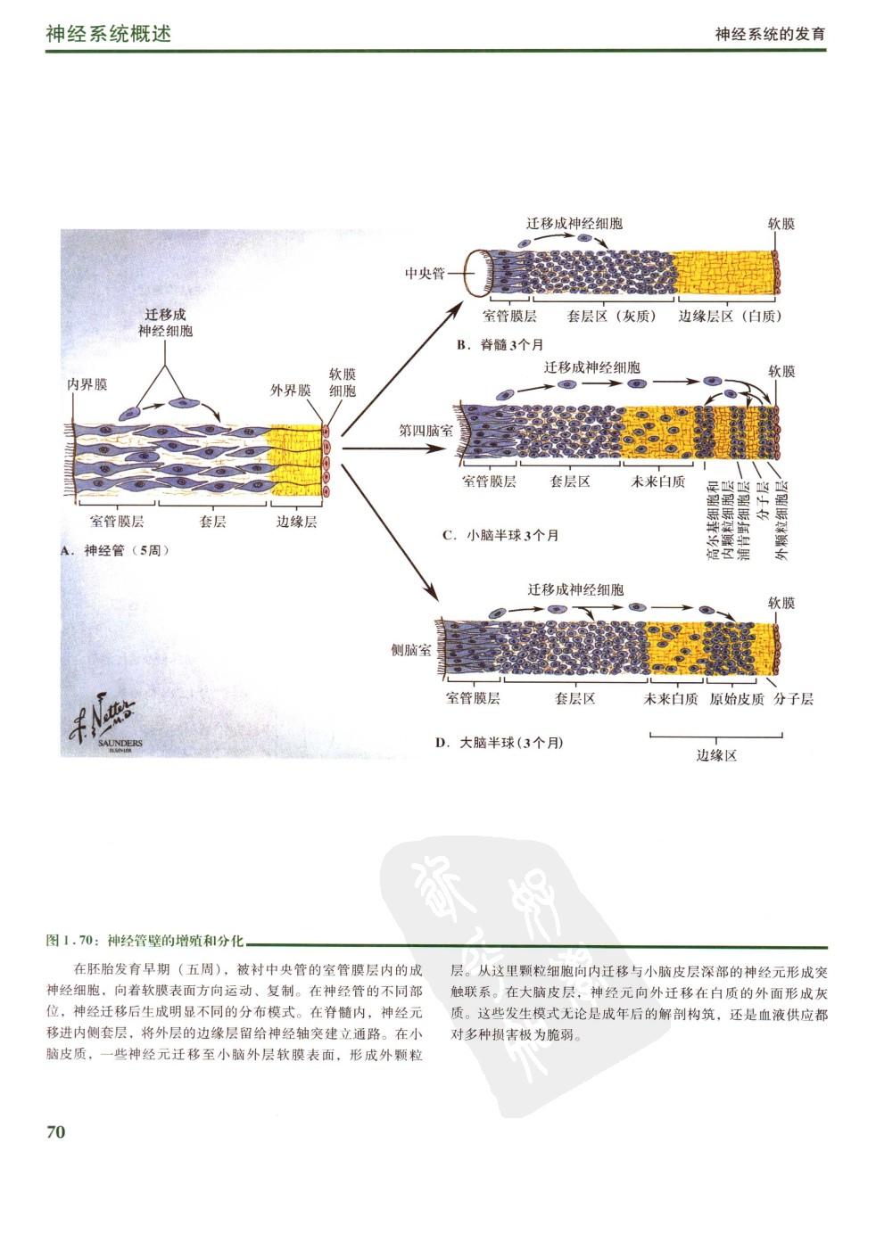
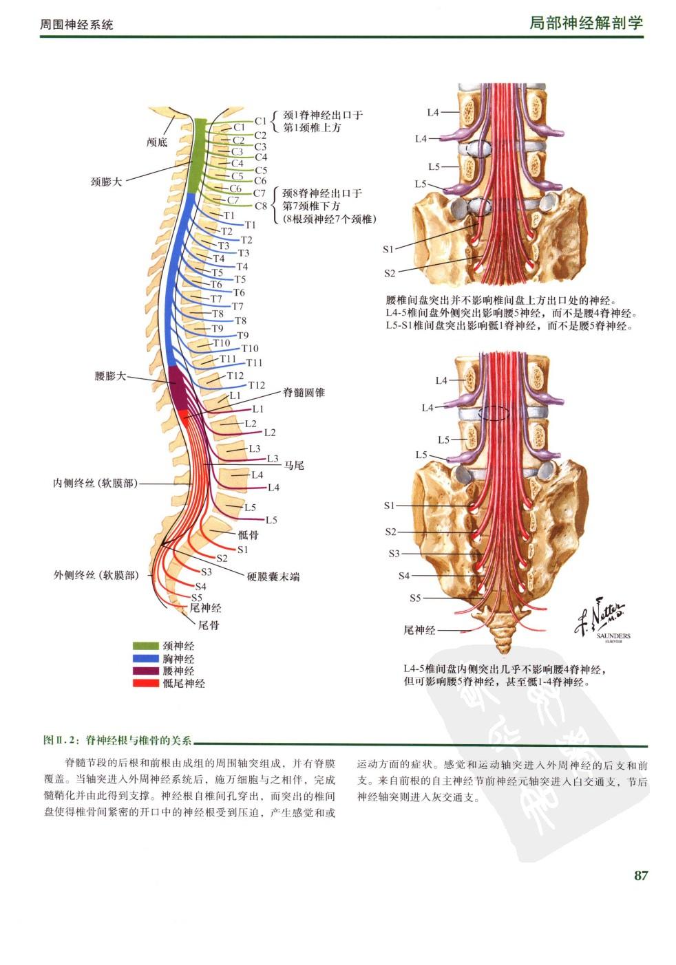
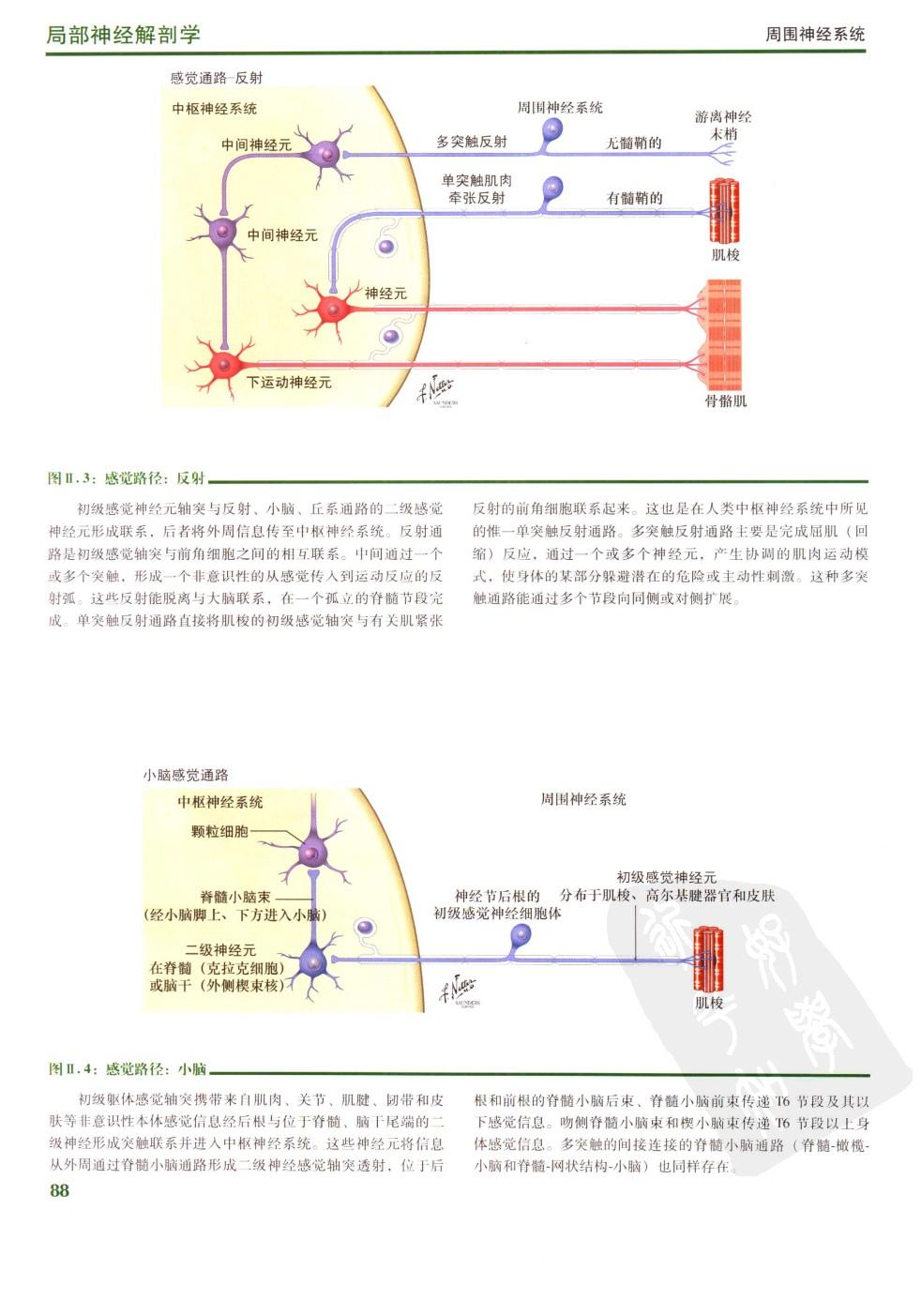
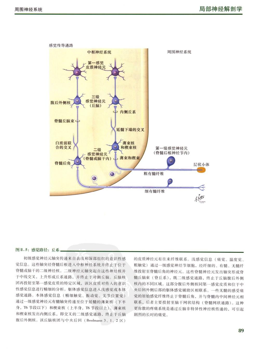
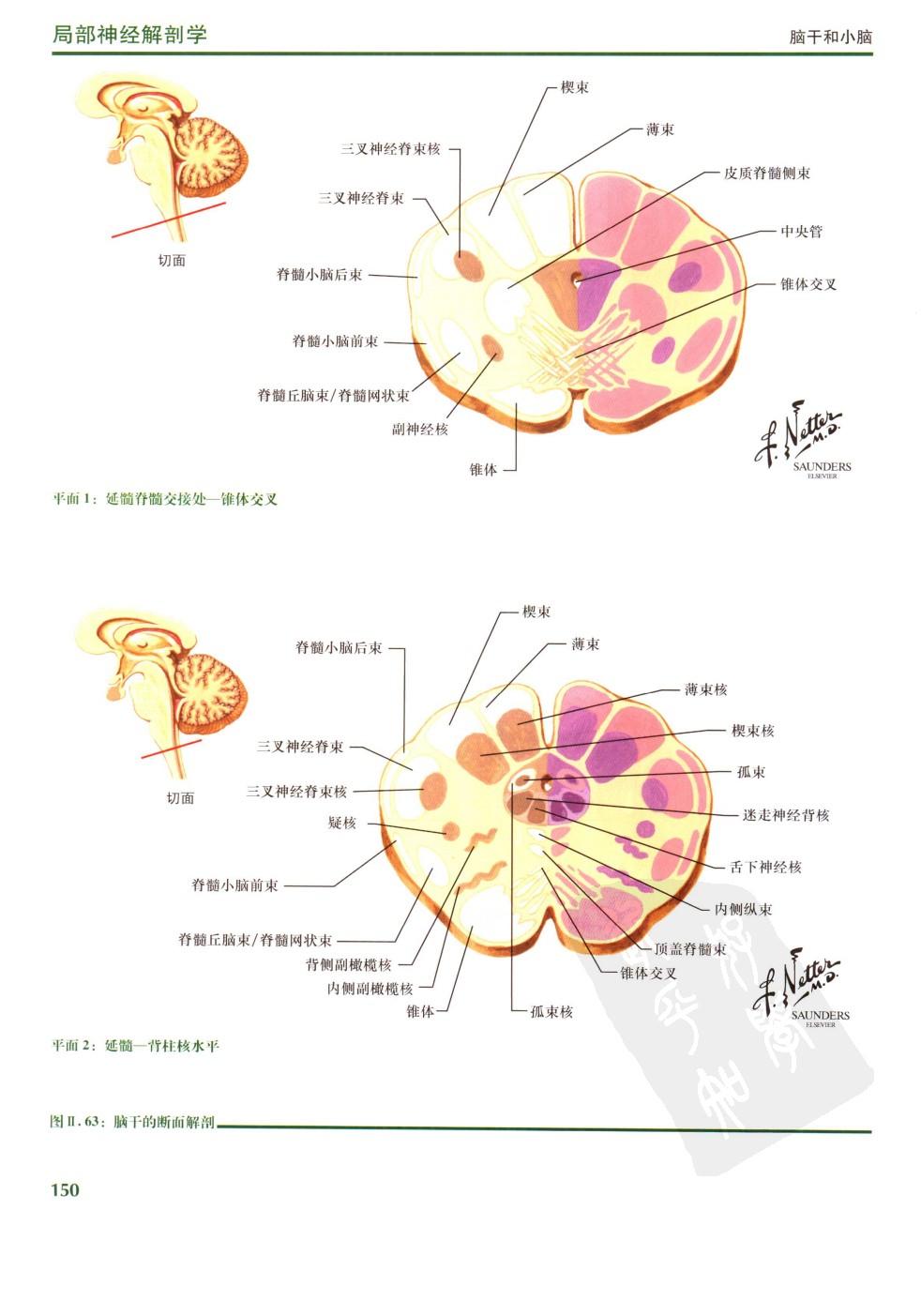
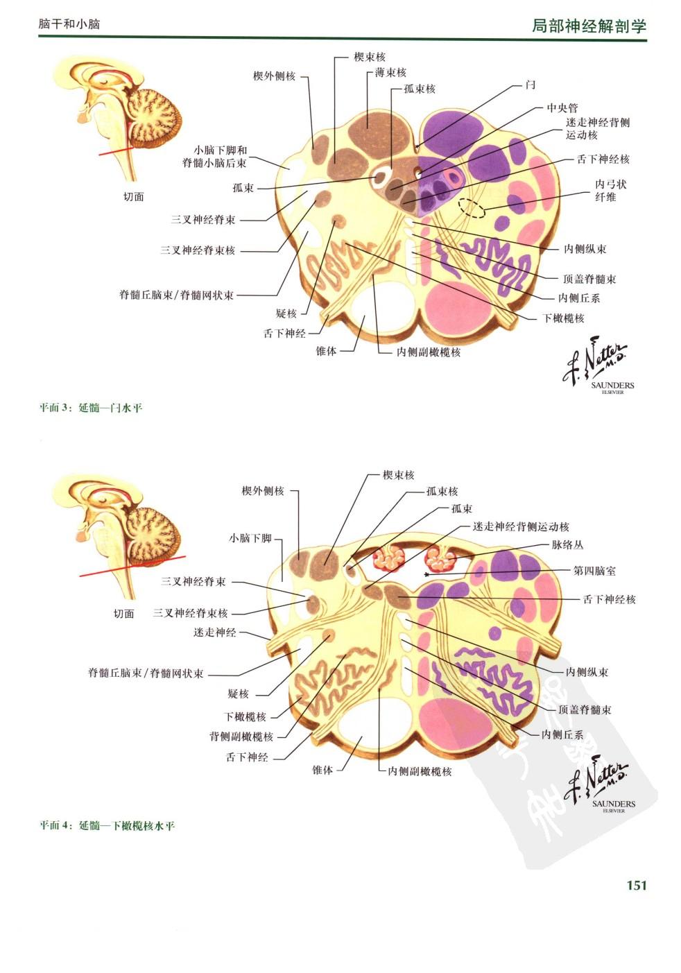
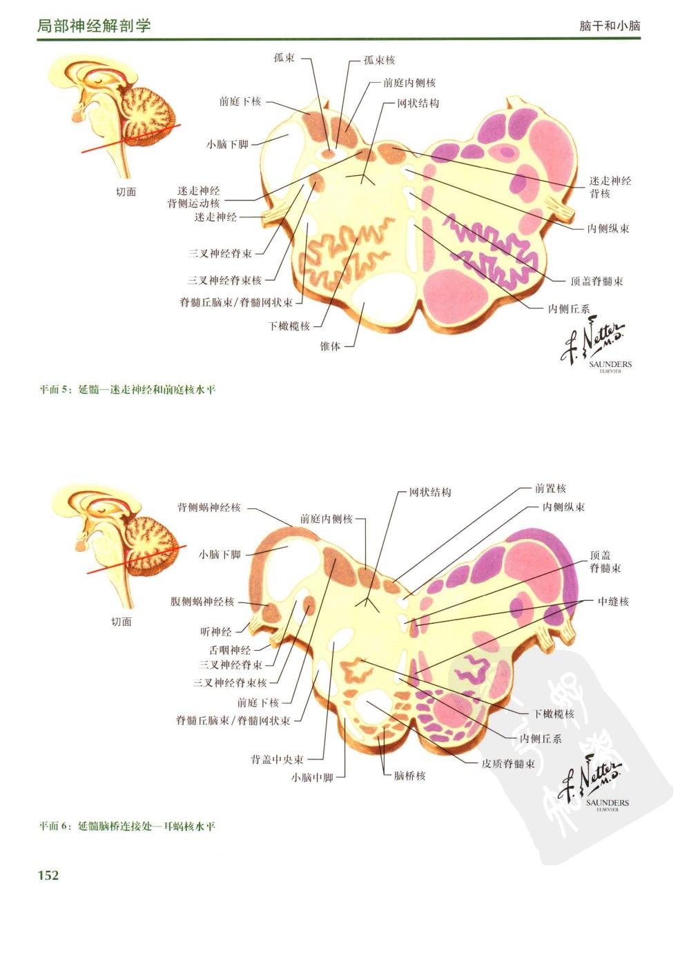
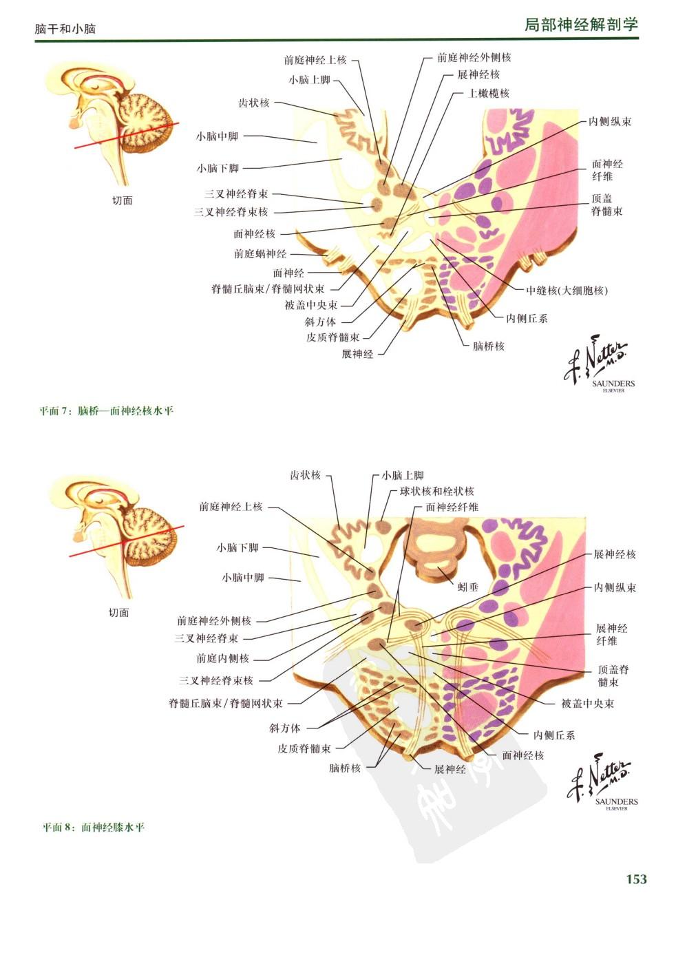
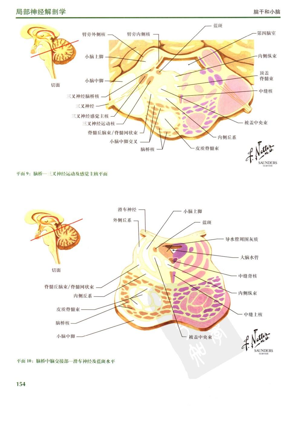
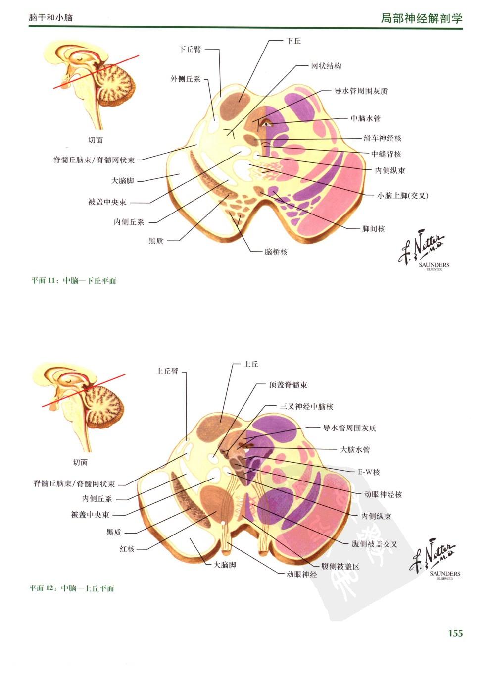
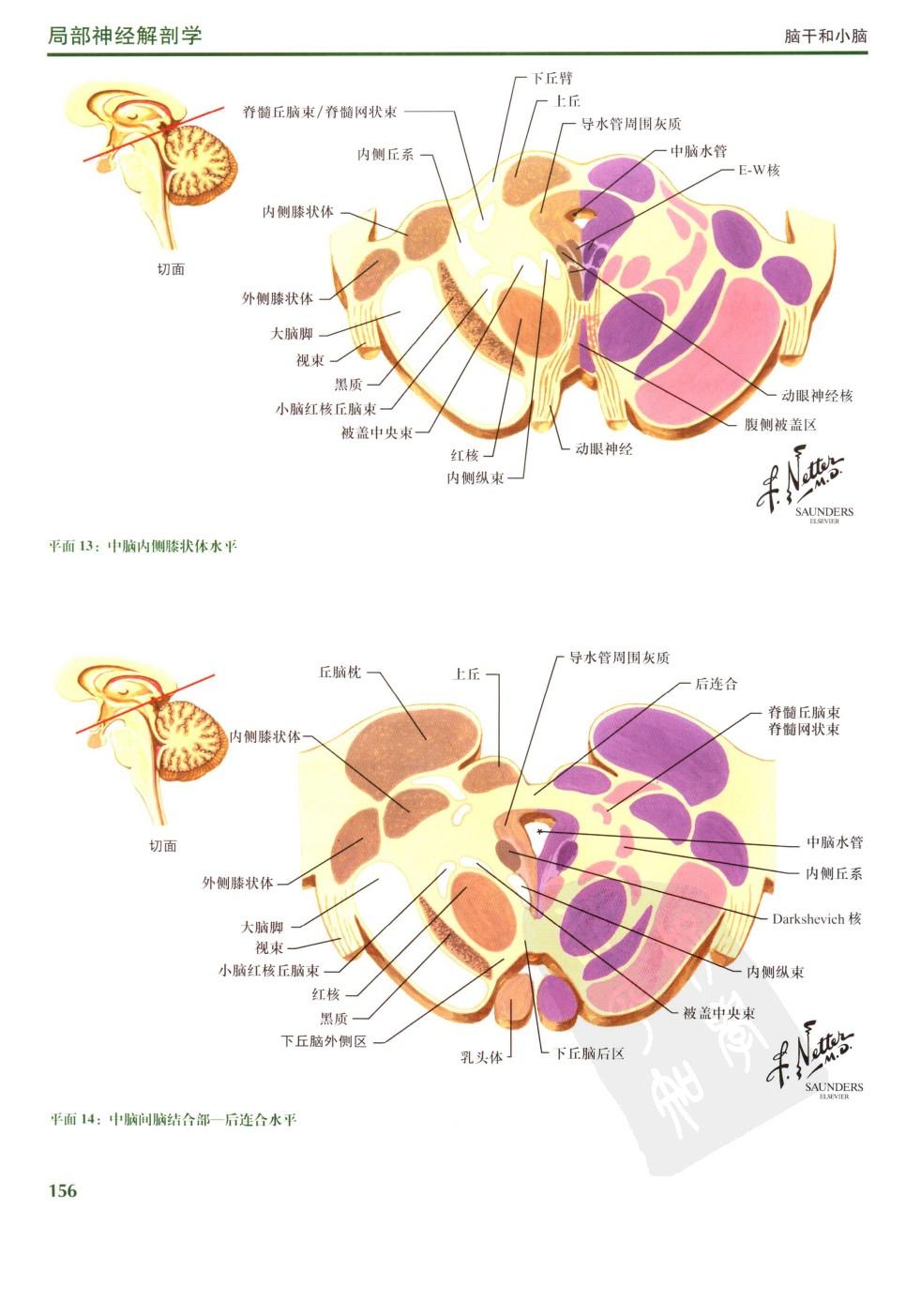
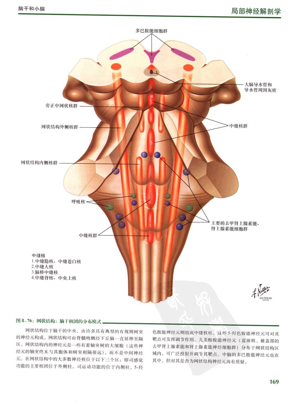
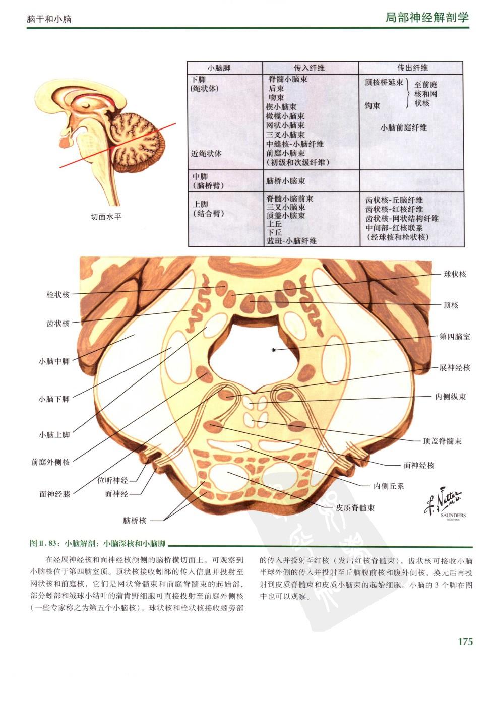
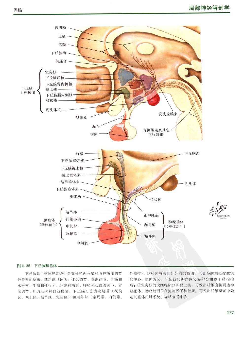
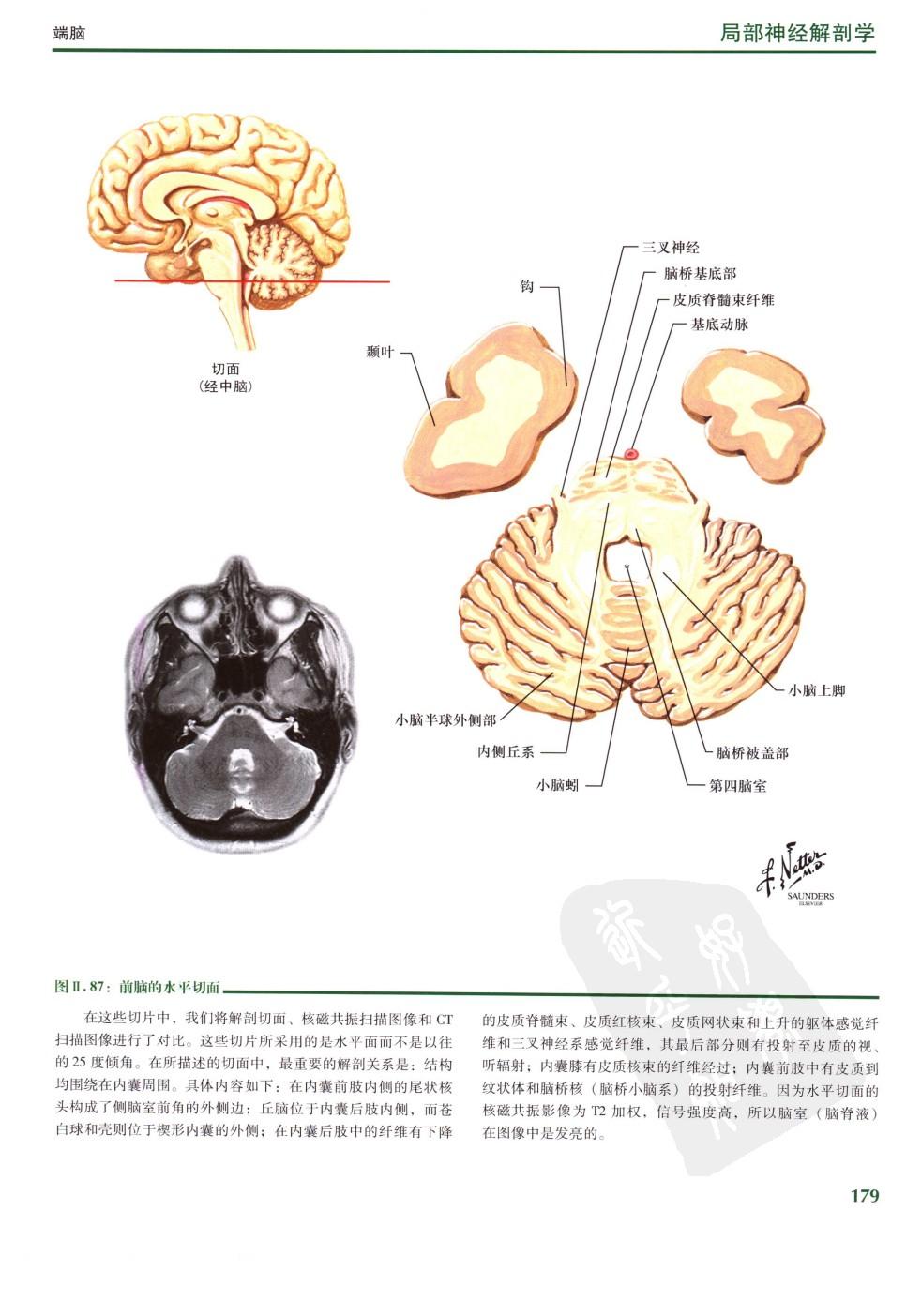
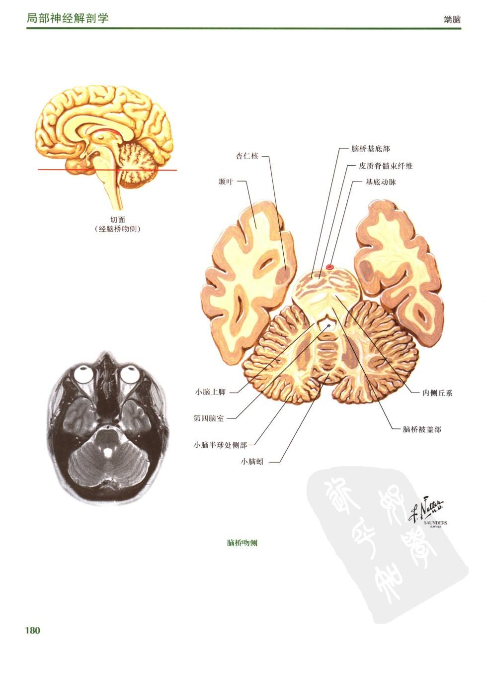
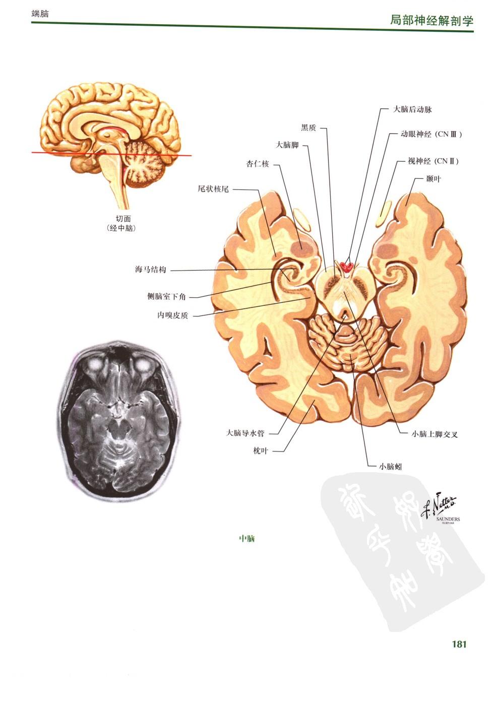
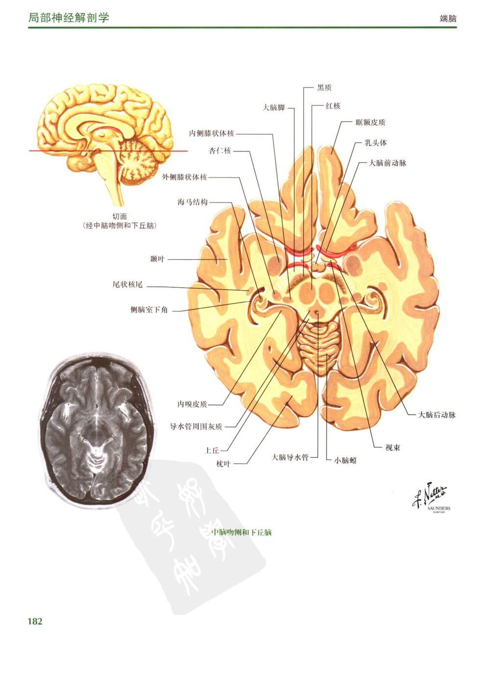
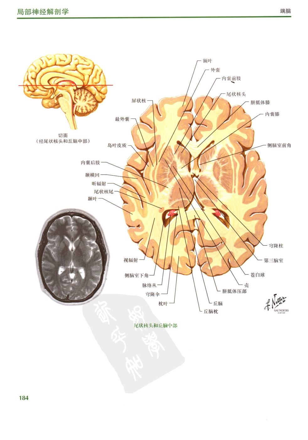
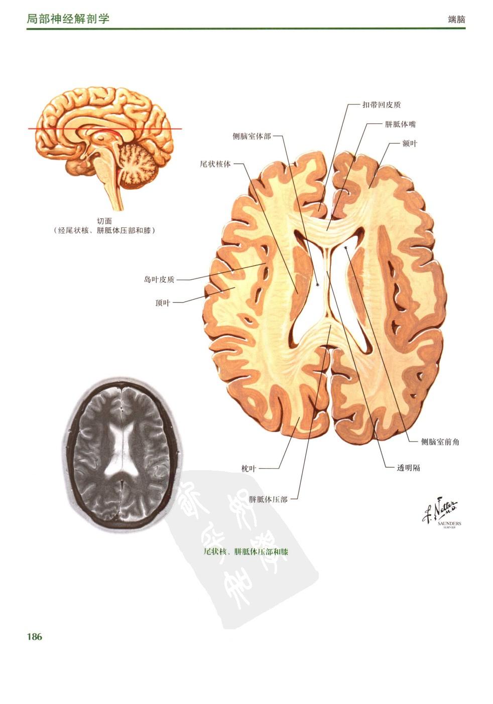
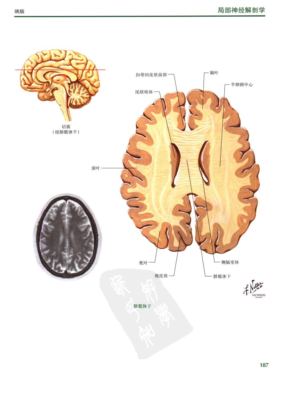
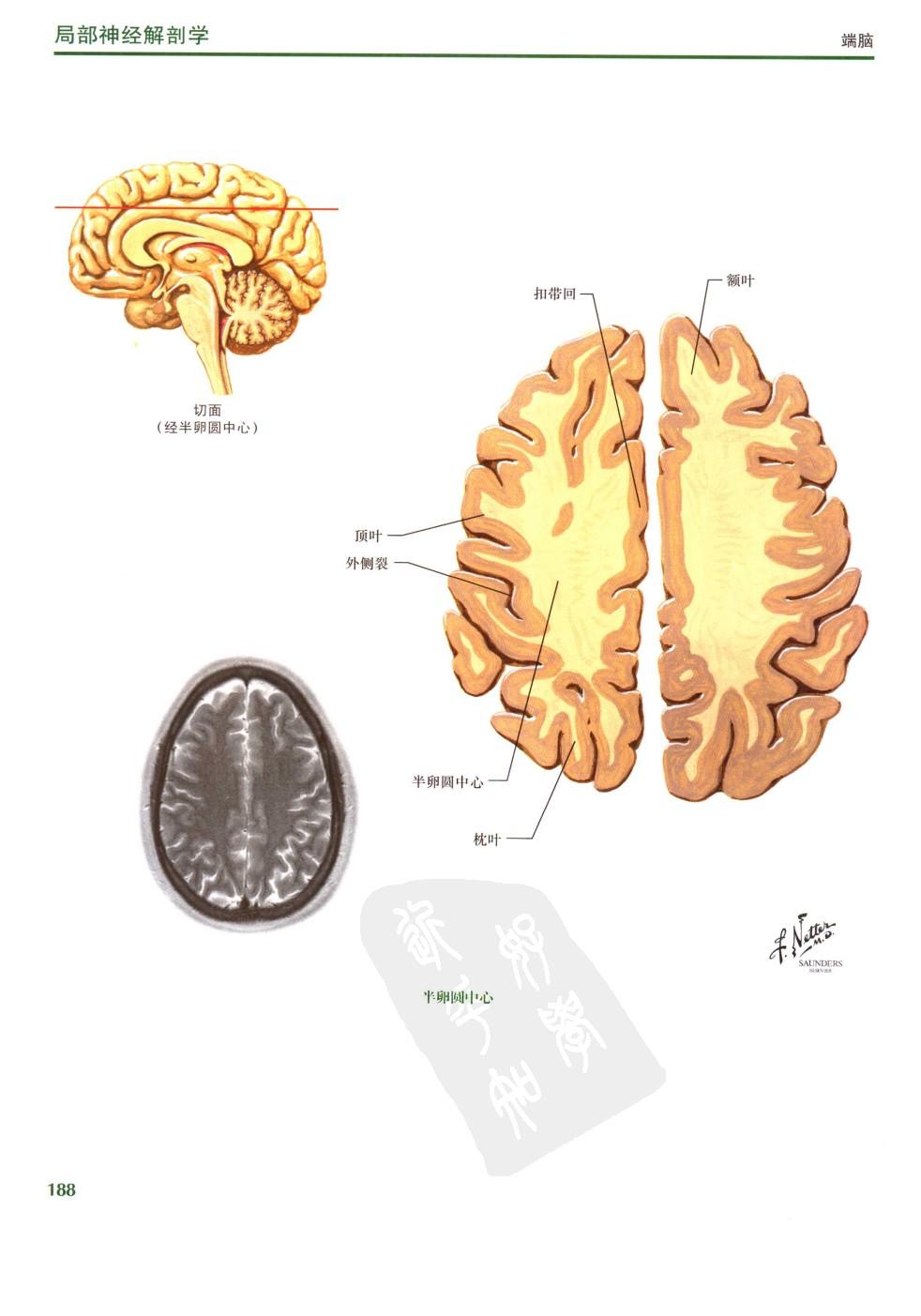
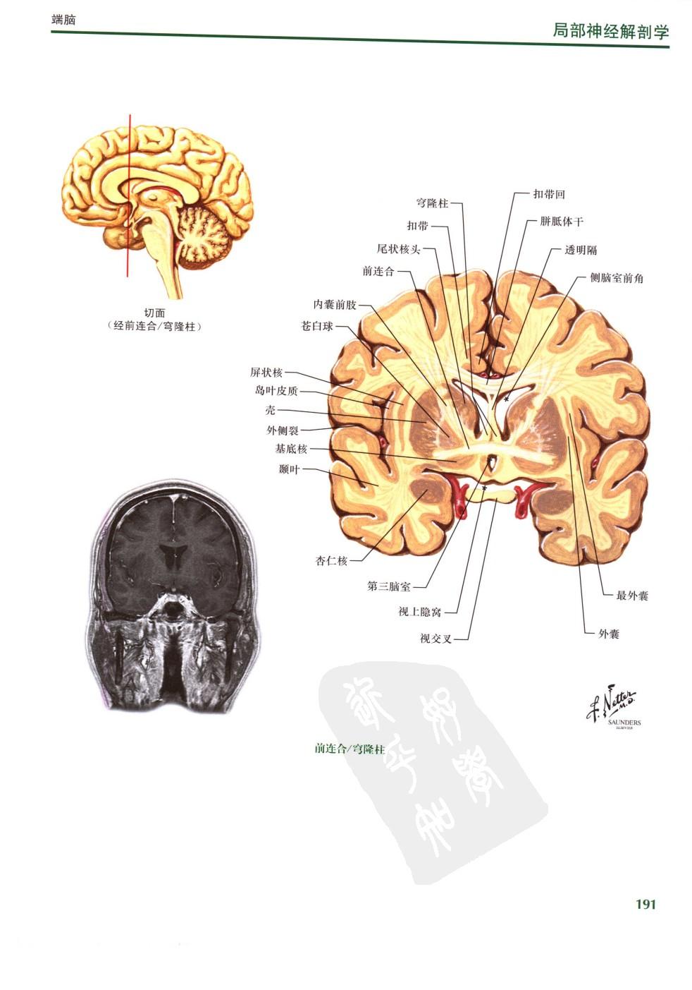
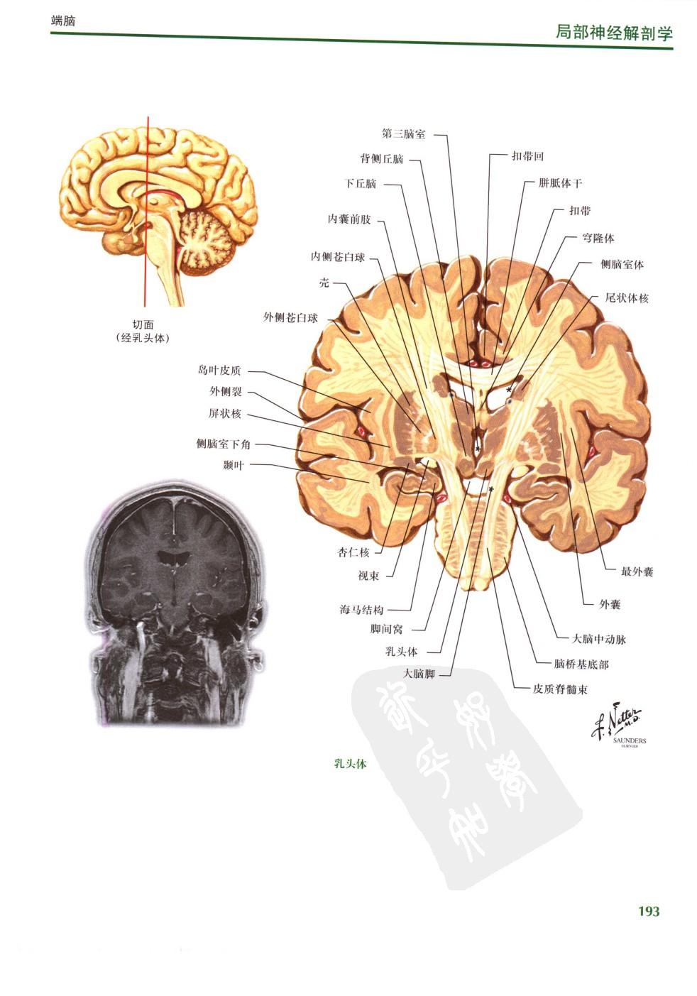
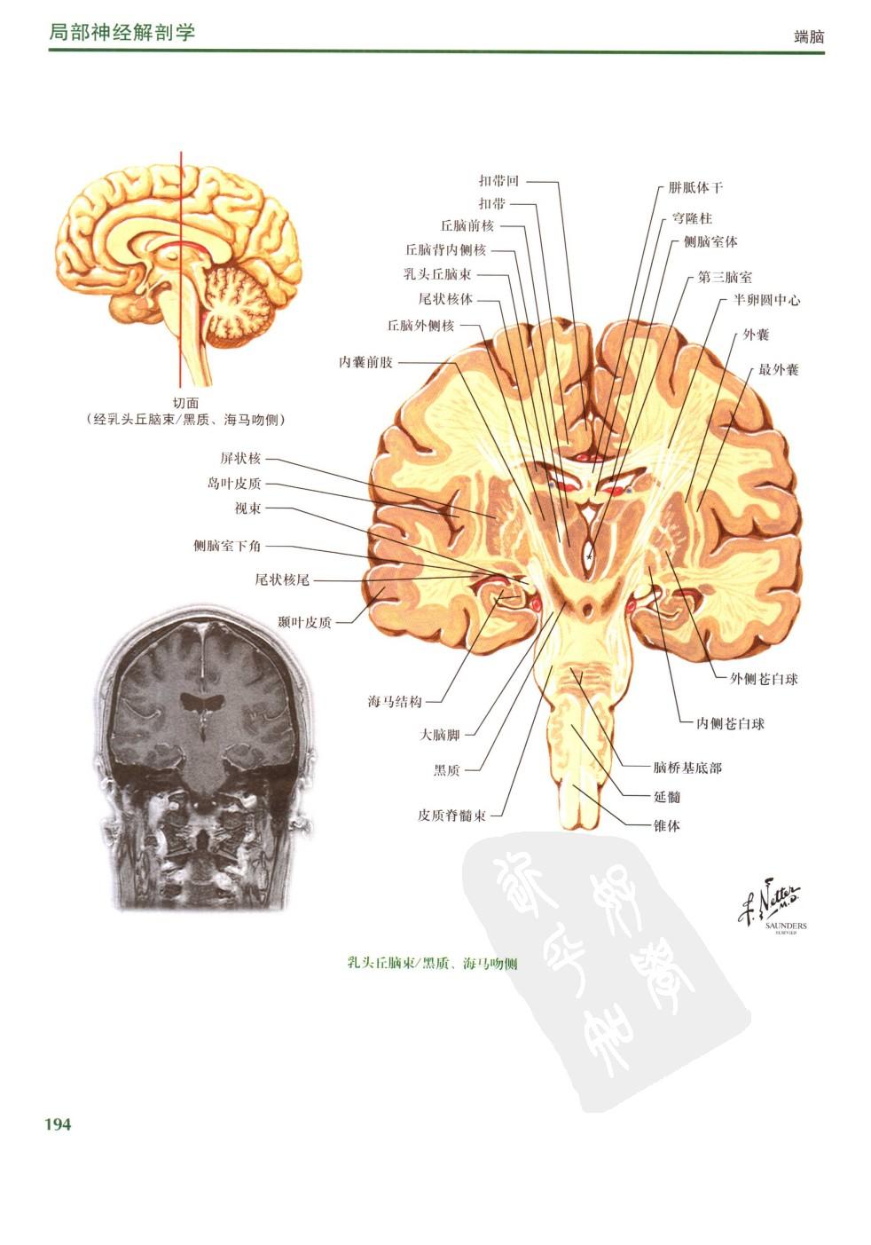
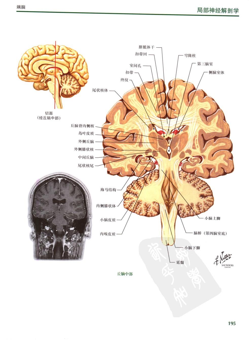
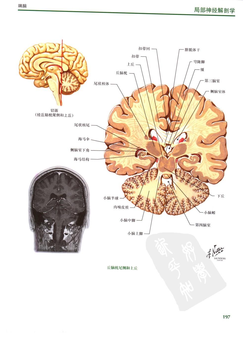
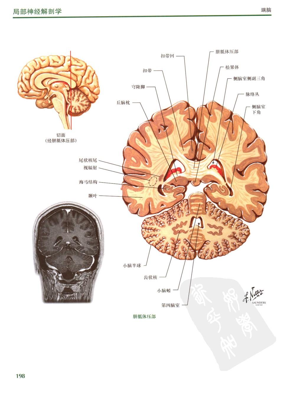
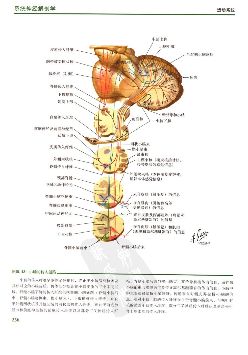
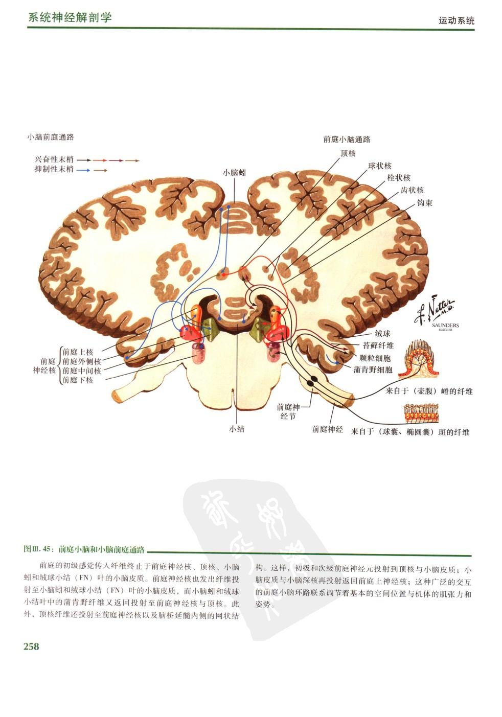
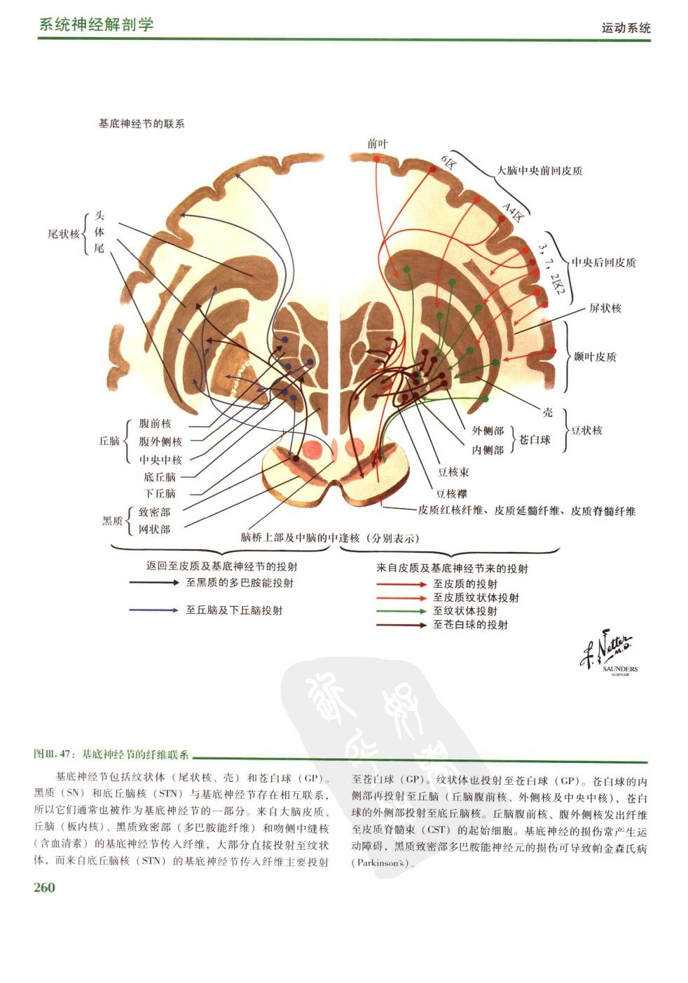
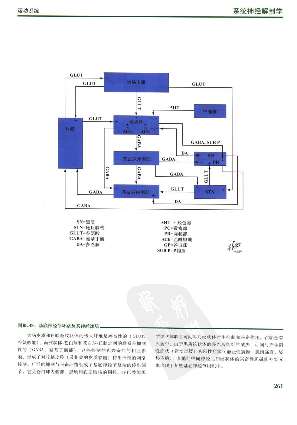
本书还通过CT、MRI的方法对脊髓和脑干作了横切、矢状与冠状扫描,并对照插图进行重点学习。与这些区域相关的详细文字描述可参阅有关神经解剖学教科书。我们用简明清晰的图标和文字介绍每张插图的主要部分和要点,特别为临床医师提供了在诊治神经科病人时所涉及的与临床相关的基础部分。通过这些精美插图并结合简洁的图标与文字描述,本书展示了局部和系统神经系统的基本成分、组成和功能。
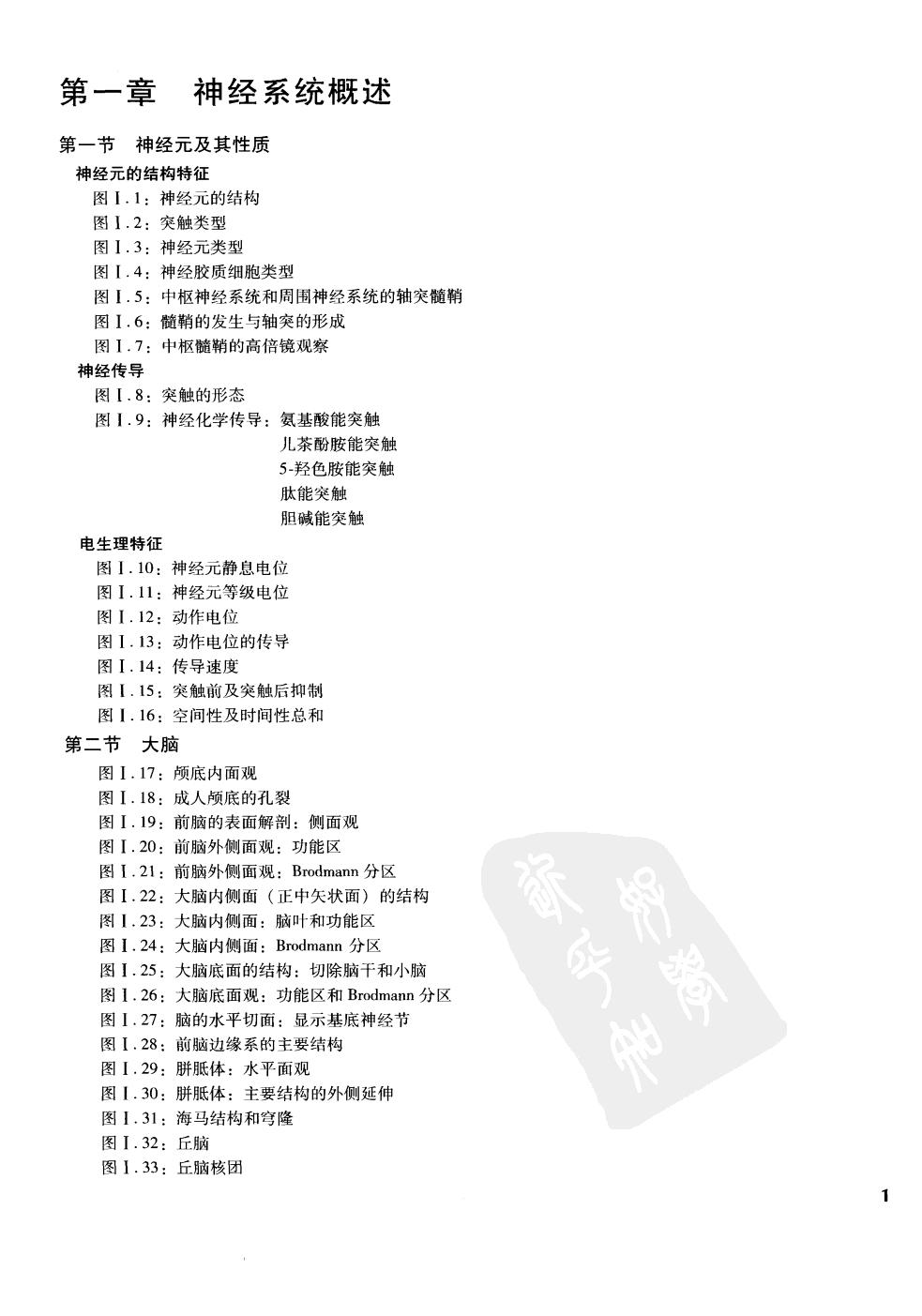
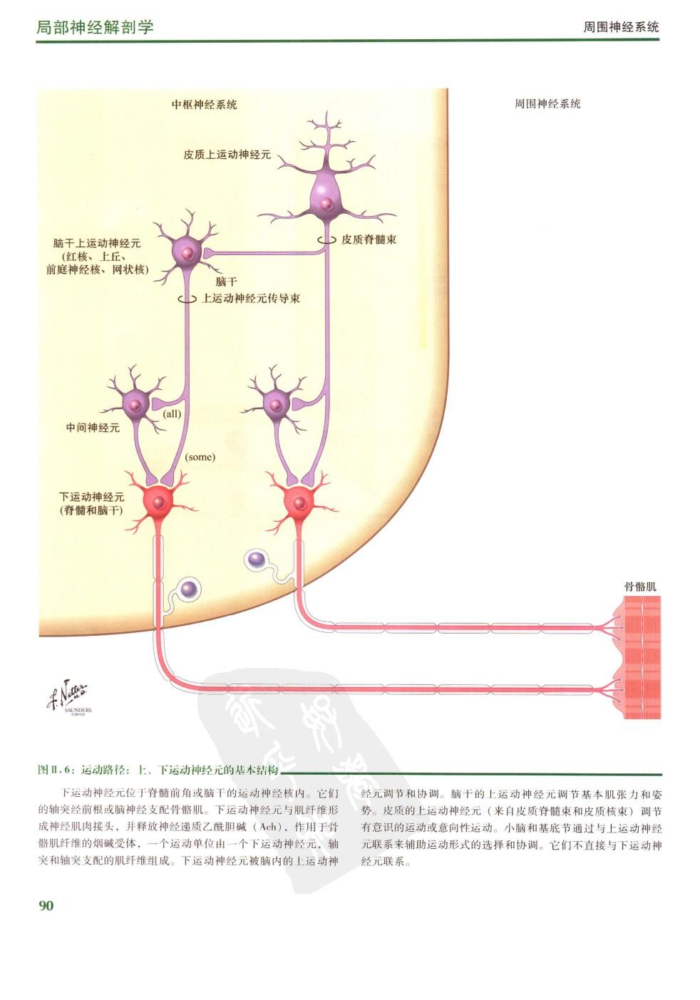
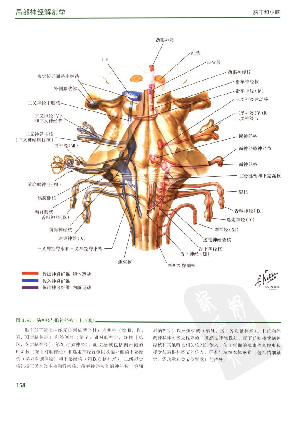
为了便于学习神经科学的学生使用本图谱,将该书分成神经系统概述、局部神经解剖学、系统神经解剖学三章予以介绍。这样的编排使读者易于通过上、下文或不同的视角来观察神经系统的复杂结构:既可观察神经系统整体的一部分,也强调了局部关系,还可以通过观察穿行于脑脊髓的某条神经系来了解其功能。掌握神经系统的基础知识使其服务于临床,为临床医师诊治神经科病人时提供帮助是本书的目标。因此以全新的观点认真学习是实现该目标有用的方法。此外,本书掌握上运动神经元与下运动神经元的构成和联系就迈向了理解I临床运动神经元疾病的第一步。进而准确定位。本书的核心内容以及将局部神经解剖和功能相互紧密联系使得学习更加容易而不是靠死记硬背。神经科学是一门十分复杂的学科,对神经系统主要区域和结构的真正理解对于基础研究与临床应用是至关重要的。
本书描绘了人体神经系统基本结构,不包括,临床神经疾病,如多发性硬化、早老性痴呆、卒中、脊髓与脑损伤、认知障碍、失用、失语等。
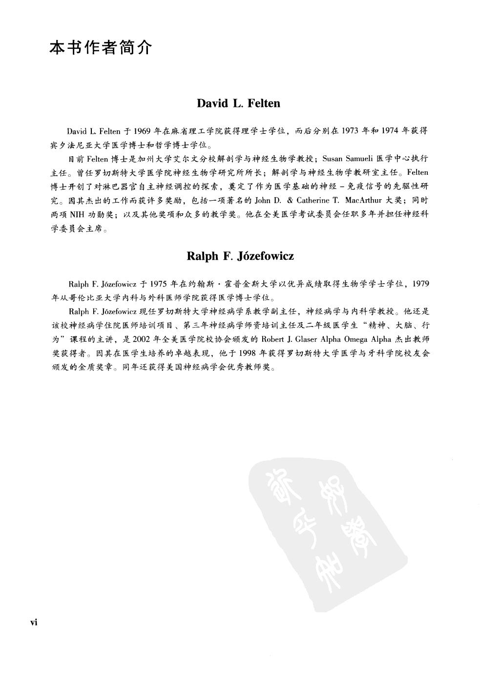
奈特博士于1906年生于美国纽约市。他曾在学生艺术联合会和美国国家设计院学习绘画艺术,后进入纽约大学医学院学习医学,于1931年获得医学博士学位。在学习期间,他的素描就引起了医学界的注意,并纷纷聘请他为一些文章和著作绘制插图。在1933年成为职业外科医生后,奈特继续在业余时间从事绘画工作,但他最终放弃了医生的职业,全身心地投入到钟爱的绘画艺术中。在第二次世界大战期间,他在美国军队服役,退役后便开始了与cIBA制药公司(现为Novartis制药公司)的长期合作。长达45年的合作使他积累了宝贵的医学艺术财富,成为世界各国的医生和其他医务工作者十分熟悉的医学绘画艺术家。
2000年7月,Icon公司获得了奈特博七的同集,并根据新的资料对奈特博士的原作不断进行修正,并增补一些新的插图,而这些插图都是由接受过奈特博士风格训练的画家所制作的。
奈特博士的作品是用图画形象地传授医学知识的典范。13卷《奈特医学图集》收入了奈特博士创作的2
“Nate Color Atlas of Human Neuroanatomy” is a book published by the People’s Health Publishing House in September 2006. The author is Felteng. This book describes the increasingly in-depth research results of many regions of the human brain, spinal cord, and peripheral nervous system.
The book “Knight’s Color Map of Human Neuroanatomy” not only covers all of Dr. Frank H. Netter’s original rich and exquisite drawings of regional and systemic neuroanatomy, but also introduces the increasingly in-depth research on many regions of the human brain, spinal cord, and peripheral nervous system, providing researchers and teachers in the field of neurology with broader and more comprehensive knowledge.
This book also uses CT and MRI methods to perform transverse, sagittal, and coronal scans of the spinal cord and brain stem, and focuses on learning by comparing the illustrations. Detailed textual descriptions of these regions can be found in textbooks on neuroanatomy. We use concise and clear icons and text to introduce the main parts and key points of each illustration, especially providing clinicians with clinically relevant basic parts involved in the diagnosis and treatment of neurological patients. Through these exquisite illustrations, combined with concise icons and textual descriptions, this book demonstrates the basic components, composition, and functions of the local and systemic nervous system.
In order to facilitate the use of this atlas by students studying neuroscience, the book is divided into three chapters: Overview of the nervous system, Regional Neuroanatomy, and Systematic Neuroanatomy. This arrangement makes it easy for the reader to observe the complex structure of the nervous system from different perspectives, such as above, below, or from different perspectives: one can observe a part of the nervous system as a whole, emphasizing local relationships, and one can also understand its function by observing a certain nervous system running through the brain and spinal cord. The goal of this book is to master the basic knowledge of the nervous system so that it can serve the clinic and provide assistance to clinicians when diagnosing and treating patients in the department of neurology. Therefore, serious learning from a new perspective is a useful way to achieve this goal. In addition, mastering the composition and connection of upper and lower motor neurons in this book is the first step towards understanding clinical motor neuron diseases. And accurate positioning. The core content of this book and the close connection between local neural anatomy and function make learning easier rather than relying on rote memorization. Neuroscience is a very complex discipline, and a true understanding of the main regions and structures of the nervous system is crucial for basic research and clinical applications.
This book describes the basic structure of the human nervous system, excluding clinical neurological diseases such as multiple sclerosis, Alzheimer’s disease, stroke, spinal cord and brain injury, cognitive impairment, aphasia, and so on.
Dr. Knight was born in New York City in 1906. He studied the art of painting at the Student Art Federation and the National Academy of Design in the United States, and then entered the Medical School of New York University to study medicine. In 1931, he received a medical doctor’s degree. During his study, his sketches attracted the attention of the medical profession, and he was hired to draw illustrations for some articles and works. After becoming a professional surgeon in 1933, Knight continued to engage in painting in his spare time, but ultimately gave up his career as a doctor and devoted himself to his beloved art of painting. During World War II, he served in the United States military, and after his retirement, he began a long-term collaboration with cIBA Pharmaceuticals (now Novartis Pharmaceuticals). After 45 years of cooperation, he has accumulated valuable medical art wealth and become a medical painting artist familiar to doctors and other medical workers around the world.
In July 2000, Icon Company obtained the same collection of Dr. Knight VII, and continuously revised Dr. Knight’s original work based on new information, adding some new illustrations, all of which were produced by painters trained in Dr. Knight’s style.
Dr. Knight’s work is an example of using images to impart medical knowledge. The 13 volume “Nate Medical Atlas” was included in the 2


Medicine医药 Record记录 309P 奈特人体神经解剖彩色图谱
历史上的今天 ( 18 ):
- 2024年-03月-29日:N64:Mystical Ninja Starring Goemon 伍佑卫门桃山幕府记
- 2024年-03月-29日:N64:Mystical Ninja 2 Starring Goemon 大盗伍佑卫门2
- 2024年-03月-29日:N64:Ms. Pac-Man Maze Madness Pac 吃豆小姐 疯狂迷宫
- 2024年-03月-29日:N64:MRC-Multi Racing Championship 多赛车锦标赛
- 2024年-03月-29日:N64:Mortal Kombat Trilogy 真人快打三部曲
- 2024年-03月-29日:N64:Mortal Kombat Mythologies: Sub-Zero 真人快打 神话 绝对零度
- 2024年-03月-29日:N64:Mortal Kombat 4 真人快打4
- 2024年-03月-29日:N64:Morita Shougi 64 N64森田将棋
- 2024年-03月-29日:N64:Monster Truck Madness 64 疯狂怪物卡车64
- 2024年-03月-29日:N64:Monopoly 强手棋
- 2024年-03月-29日:N64:Monaco Grand Prix – Racing Simulation 2 模拟赛车2-摩纳哥大奖赛
- 2024年-03月-29日:N64:Mission Impossible 碟中谍
- 2024年-03月-29日:Culture文化:玲珑1931_020 (39P)
- 2024年-03月-29日:Culture文化:玲珑1931_019 (39P)
- 2024年-03月-29日:Culture文化:玲珑1931_018 (37P)
- 2024年-03月-29日:Culture文化:玲珑1931_017 (39P)
- 2024年-03月-29日:Culture文化:玲珑1931_016 (35P)
- 2024年-03月-29日:News新闻:2024年3月29日新闻简报
可点 ➠ 2023年-03月-29日 ➠ 256 s ➠ ♥ 0